International Society of Urological Pathology
Author: Marit Bernhardt, MD, Glen Kristiansen, MD; Institute of Pathology, University Hospital Bonn, Germany
72-year-old man with a mass in pararectal/paraprostatic soft tissue

Discussion of case
Solitary Fibrous Tumor (SFT) is a soft tissue neoplasm of fibroblastic origin and uncertain clinical behavior. Originally described as a pleural tumor, it has by now been shown to occur at any site of the body, most commonly intrathoracic and intraabdominal, including retroperitoneum and pelvis 1. Rarely SFT even arises in the prostate 2. SFT may occur at any age of adulthood but typically, patients are in their 5-6th decade 1. Tumors mostly present as well demarcated lesions and show a characteristic so called “patternless pattern”. Herein, hypo- and hypercellular areas are found. Often tumor cells are separated by thick bundles of collagen. Typically, staghorn like branching vessels can be found and serve as a diagnostic clue. The fat forming type of SFT, which is uncommon, includes a variably prominent component of single to large numbers of mature adipocytes. Tumor cells show a spindled to ovoid morphology with ovoid nuclei 3. On the molecular level, SFT is characterized by a NAB2::STAT6 fusion, which leads to overexpression of STAT6 protein, that can be detected by immunohistochemistry4. Features that have been associated with potential aggressive behavior which occurs in approximately 10% of cases are large tumor diameter of more than 100 mm and increased number of mitotic figures (> 4/10 HPF) 3. In addition, higher patient age, high cellularity and nuclear pleomorphism have been associated with a higher risk of relapse. SFT is treated by excision with adjuvant radiotherapy in cases of a malignant SFT 5.
Key differential diagnosis
References
Summary of clinical history
The patient presented to the outpatient department due to PSA elevation. An MR tomography of the prostate revealed a mass, 3.8 cm in diameter, located in proximity to prostate and rectum with sharp borders.
Gross findings
Core biopsies of the prostate and the soft tissue mass were received. All cores had a grey tan color.
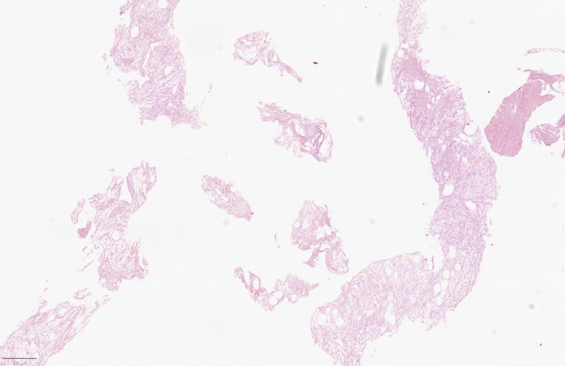
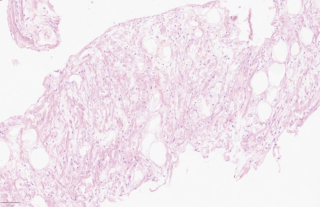
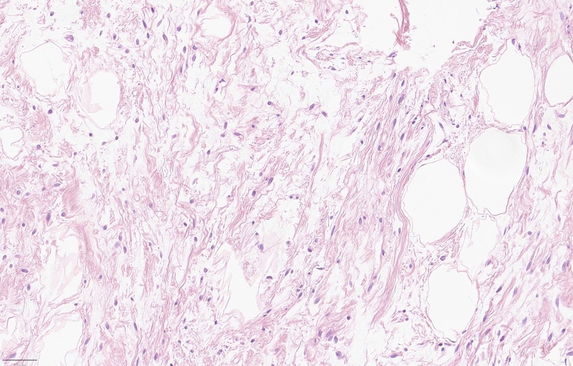
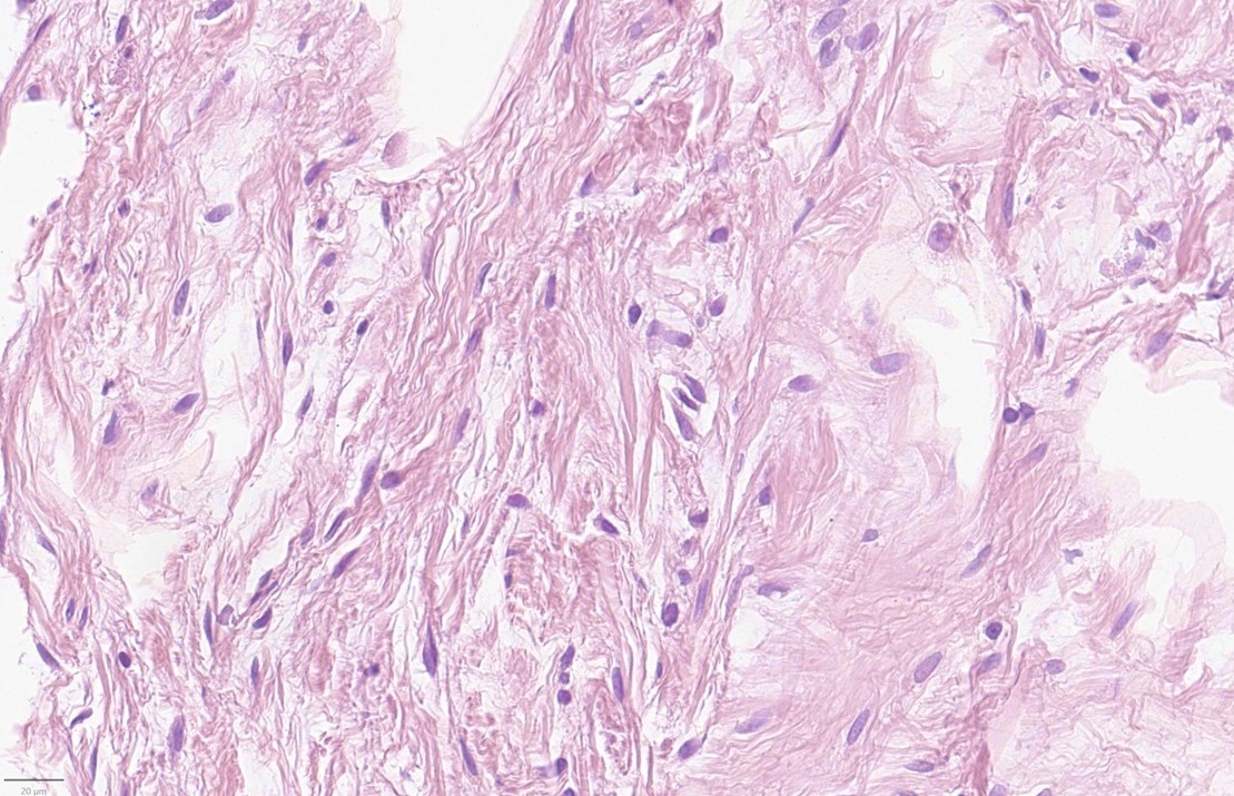
Microscopic findings
Histology showed loose soft tissue consisting of spindle cells with small round to ovoid inconspicuous nuclei. Occasional entrapment of mature adipocytes was appreciated.
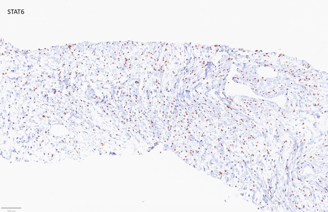
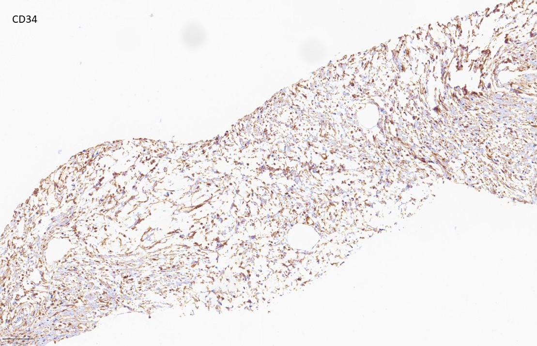
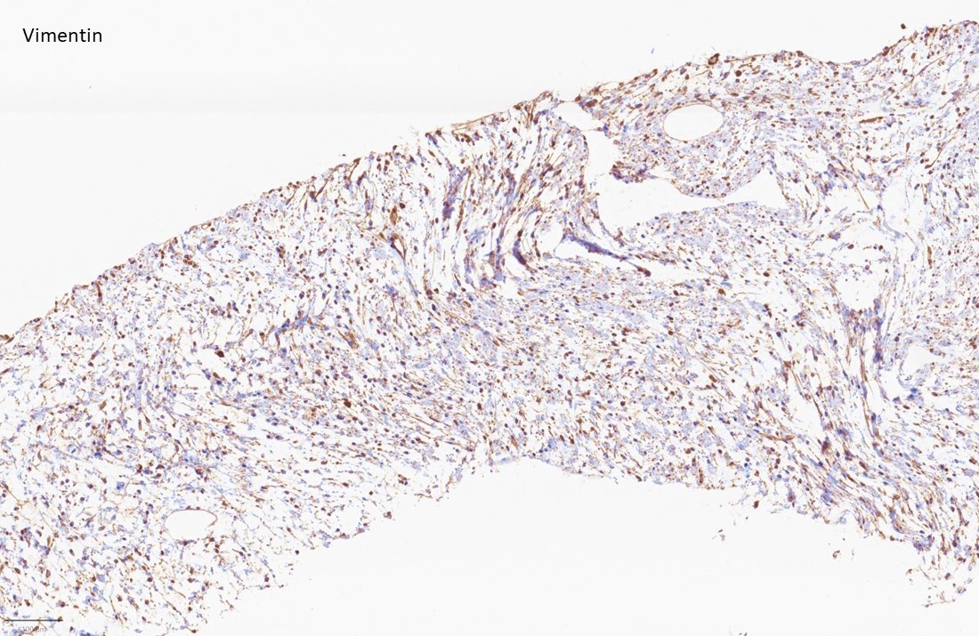
Immunohistochemical findings
The lesional cells showed a strong nuclear expression of STAT6. In addition, staining for CD34 and vimentin was positive. MUC-4, beta-Catenin and S100 were tested, too, and were negative.
CLICK IMAGE TO ENLARGE