International Society of Urological Pathology
Author: Katrina Collins, MD Indiana University School of Medicine, Indianapolis, IN, USA
53-year-old female with renal mass
Discussion of case
The main differential diagnoses in this case includes metanephric adenoma, the solid variant of papillary RCC, and adult Wilms tumor. An intriguing contradiction lies in the fact that while papillary RCC, a malignant tumor, is often encapsulated, metanephric adenoma, considered a benign tumor, typically lacks encapsulation. Wilms tumor typically exhibits two or three components—epithelial, blastema, and stromal elements—although monophasic cases are rare. Its cellular morphology appears less mature compared to metanephric adenoma, and it usually displays a higher mitotic rate.
Immunohistochemistry might prove helpful in distinguishing between metanephric adenoma and papillary RCC. Papillary RCC is positive for CK7, AMACR, and EMA, whereas metanephric adenoma usually does not show these markers. Metanephric adenoma may exhibit positivity for WT1, CD56, and CD57, while papillary RCC is usually negative for these markers. However, these immunomarkers are not useful in distinguishing metanephric adenoma from adult Wilms tumor, as they often share the same staining patterns.
Key differential diagnosis
Papillary renal cell carcinoma, particularly solid variant:
Less tightly packed tubules and less basophilic, usually has foamy macrophages
Vimentin+, PAX8+, CK7+, AMACR+, WT1-, CD57-, BRAF-
Nephroblastoma:
Difficult to differentiate if tumor is monophasic, (blastema component only)
PAX8+, WT1+, vimentin-, CD57-, BRAF-
Metanephric-like ALK rearranged renal cell carcinoma:
Similar morphology to metanephric adenoma
Vimentin+, PAX8+, CK7+, ALK+, AMACR+, BRAF-, CD57-, WT1-
Metastatic thyroid carcinoma:
Tumor cells with nuclear grooves and pseudonuclear inclusions
PAX8+, TTF1+, BRAF+, WT1-
References
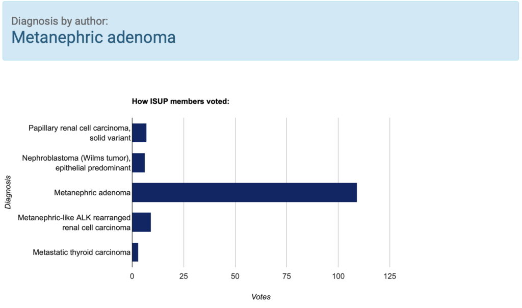
Summary of clinical history
53-year-old female with well-circumscribed right renal mass, partial nephrectomy was performed
Gross findings
4.7 cm tan, well-circumscribed and focally hemorrhagic mass, confined to kidney
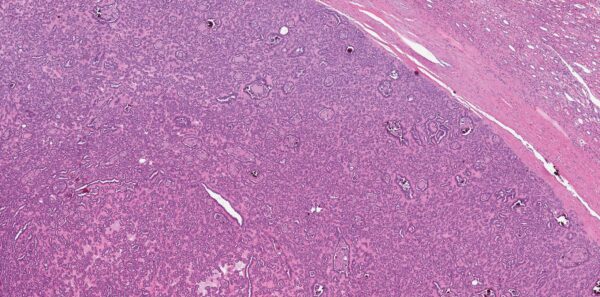
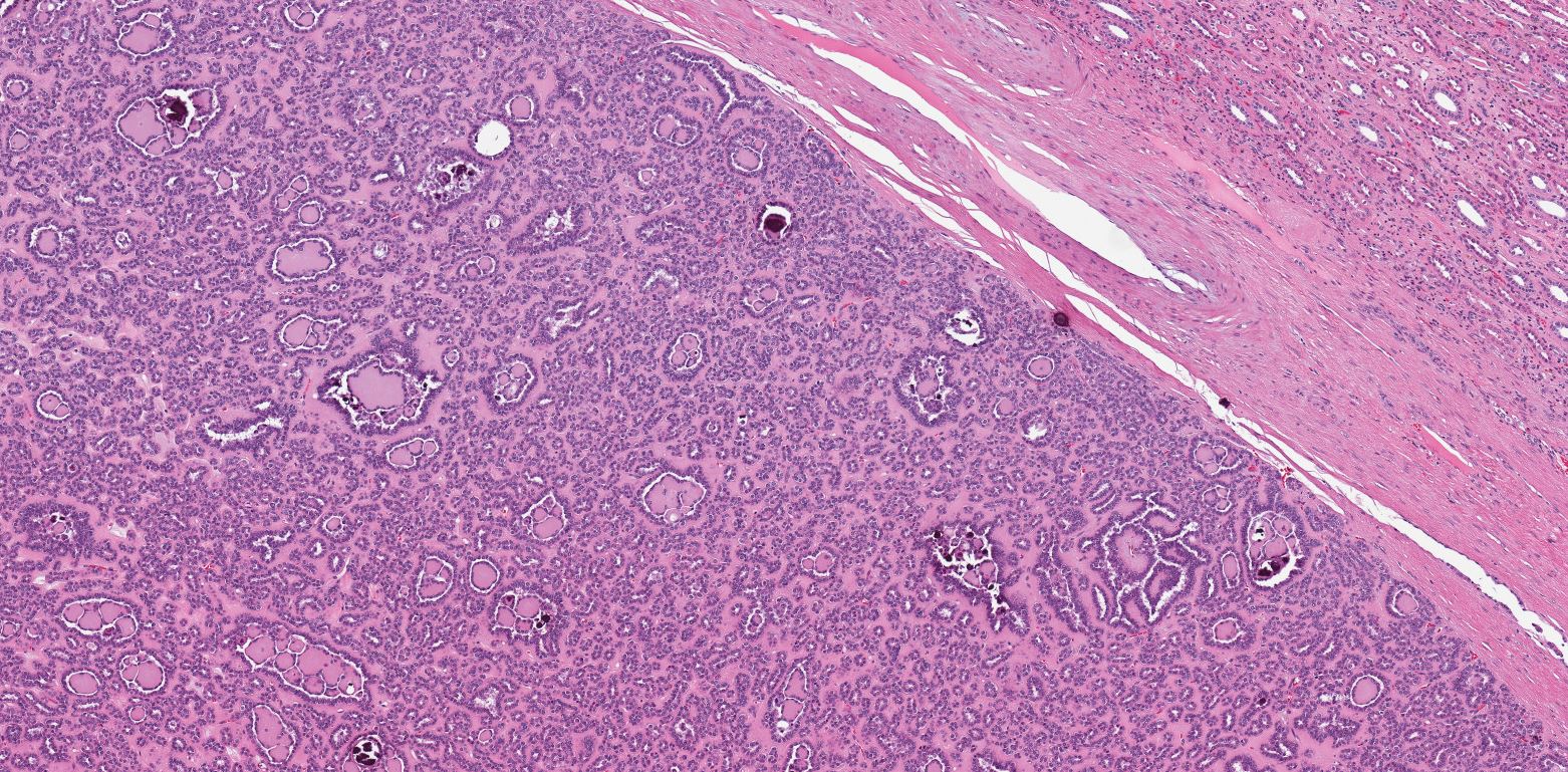
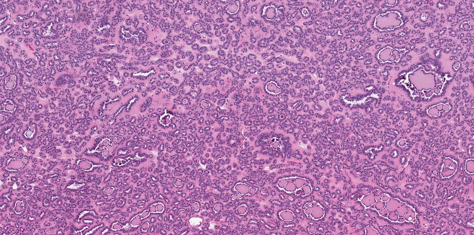
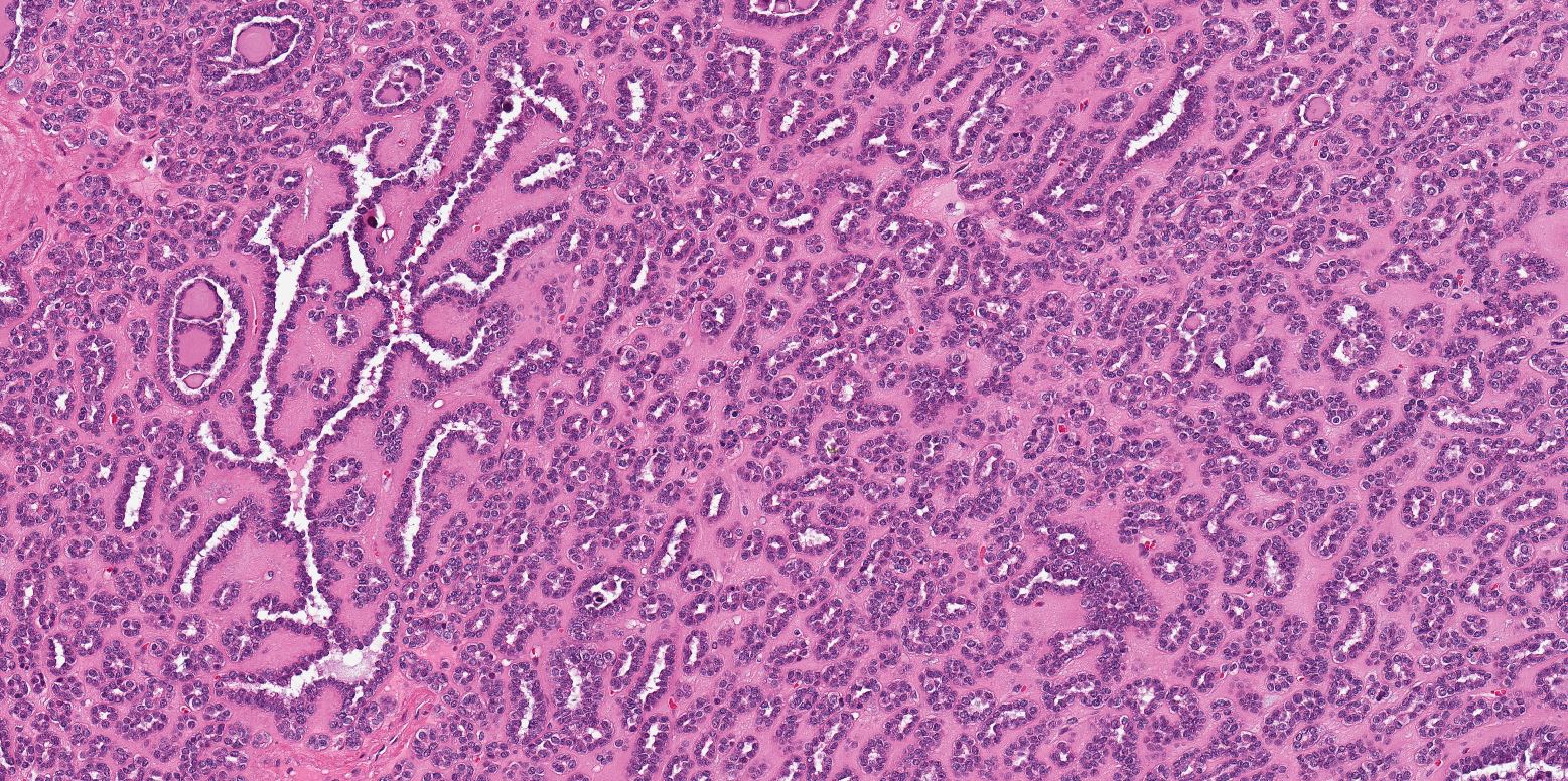
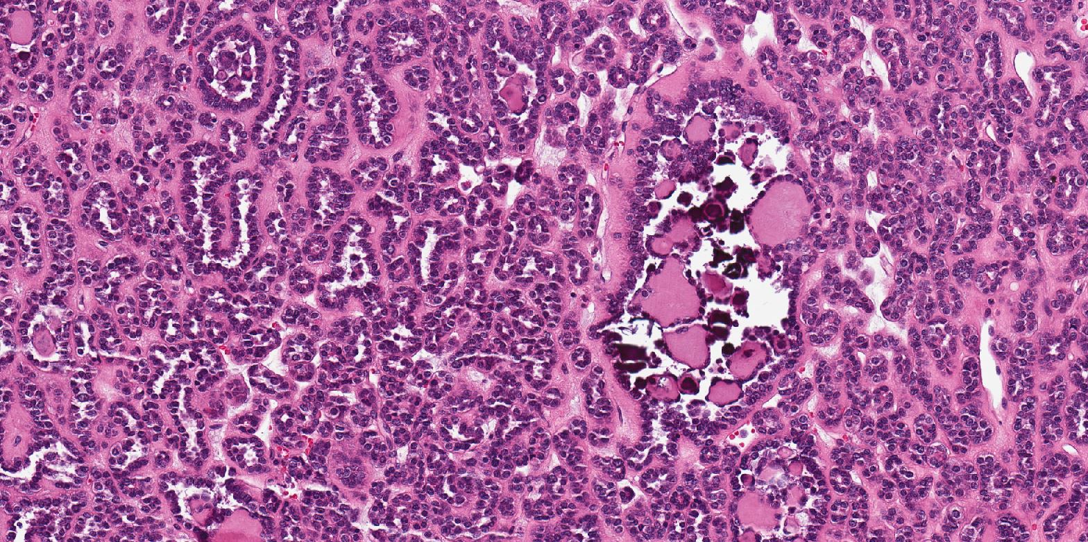
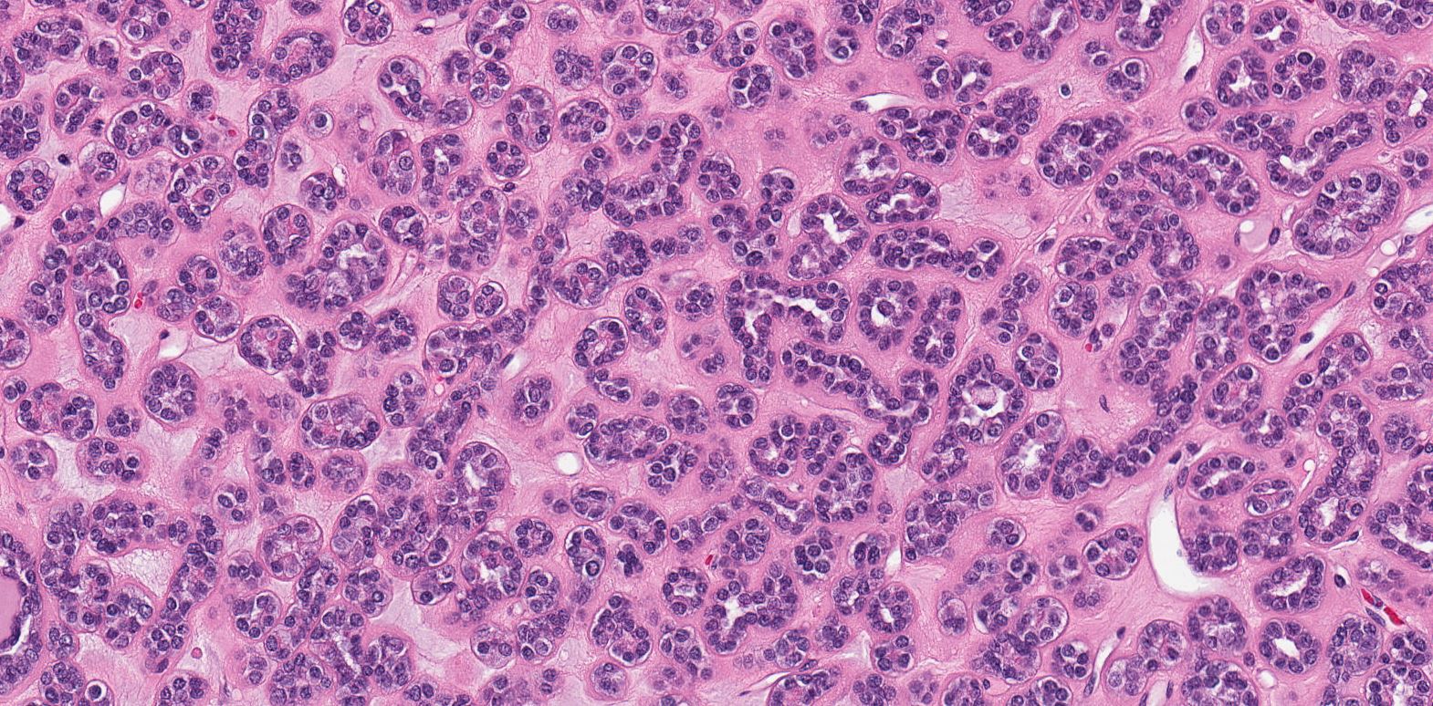
Microscopic findings
The tumor is well-circumscribed and composed of tightly packed tubules. Tumor nuclei are round or oval with occasional grooves and no obvious nucleoli are present and the cell cytoplasm is scant. In some areas, curvilinear tubules imparting papillary appearance are seen. Psammomatous calcifications are abundant.
_________________
Immunohistochemical findings
Positive: WT1, CD57, BRAF, PAX8
Negative: CK7, AMACR
CLICK IMAGE TO ENLARGE
Immunohistochemical findings
Positive: WT1, CD57, BRAF, PAX8
Negative: CK7, AMACR