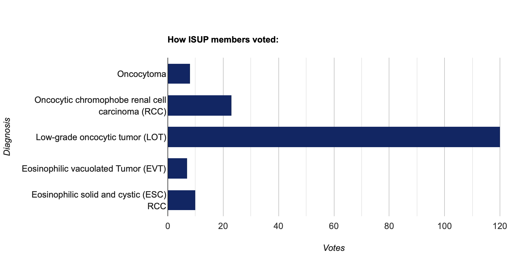
International Society of Urological Pathology
Author: Chara Ntala, Colan Ho-Yen; St George’s University Hospital, UK
62-year-old female with renal mass

Discussion of case
The landscape of renal neoplasia has significantly expanded in the past years, particularly in the field of renal oncocytic tumours. It is increasingly recognised that there are oncocytic tumours that do not fit into any of the traditional ‘eosinophilic chromophobe RCC’ and ‘oncocytoma’ tumour categories (1–3). A newly described, currently provisional entity is Low-grade oncocytic tumour (LOT) of the kidney, which shows a fairly consistent morphological and immunohistochemical profile. These mostly coccur in the sporadic setting and have very rarely been associated with tuberous sclerosis (3).
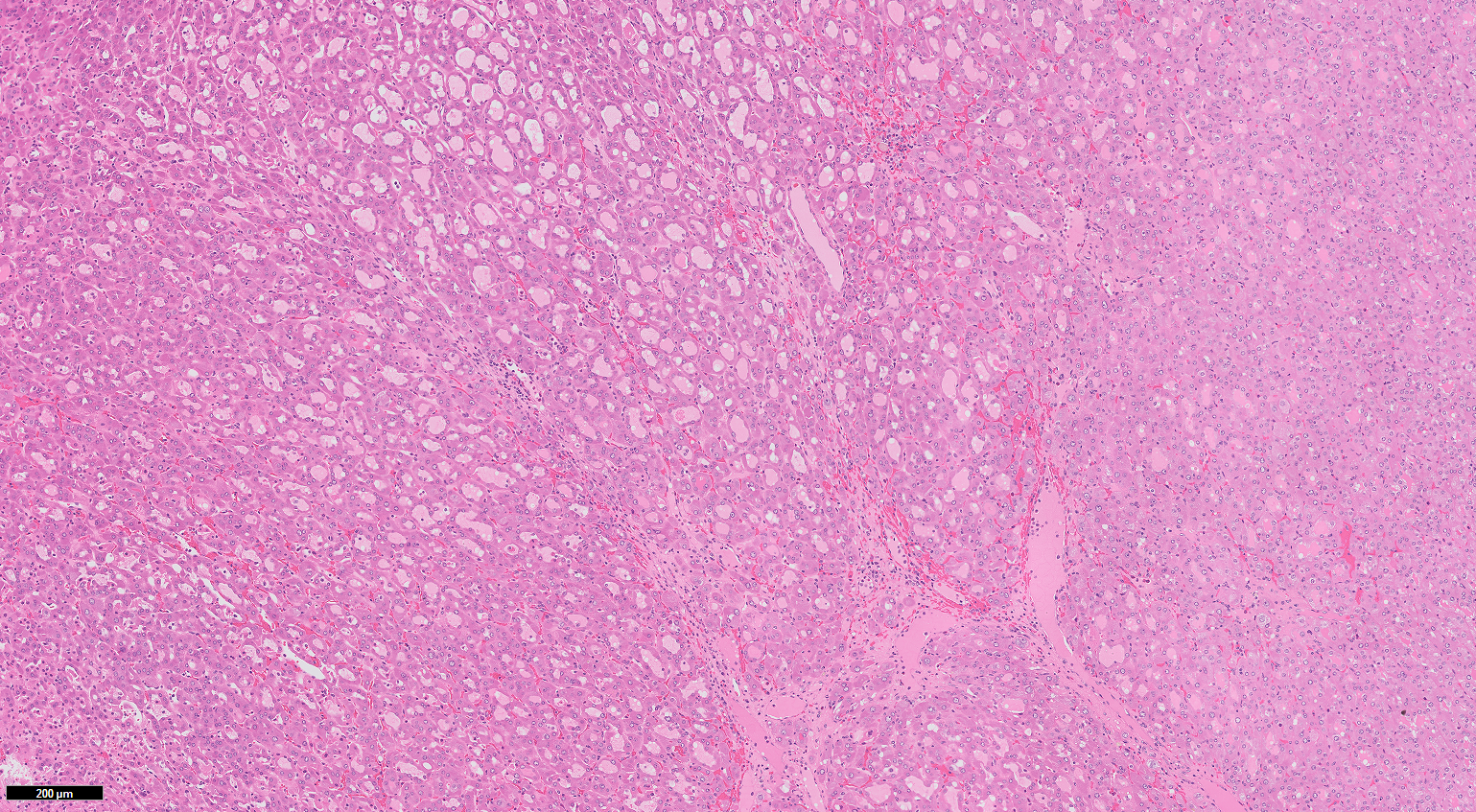
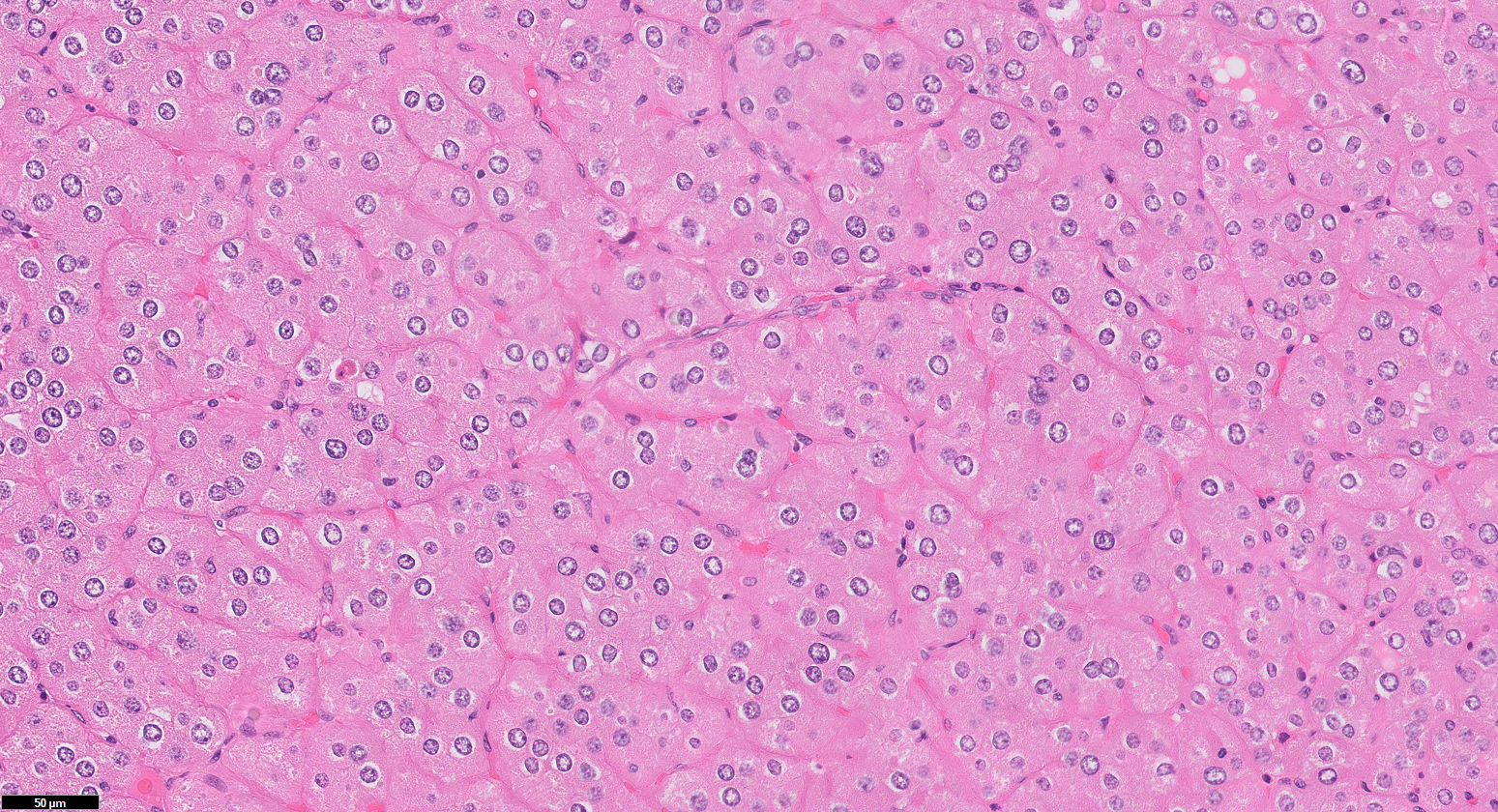
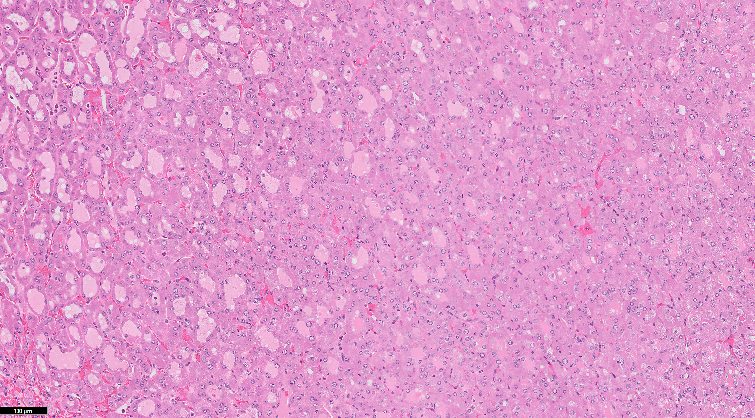
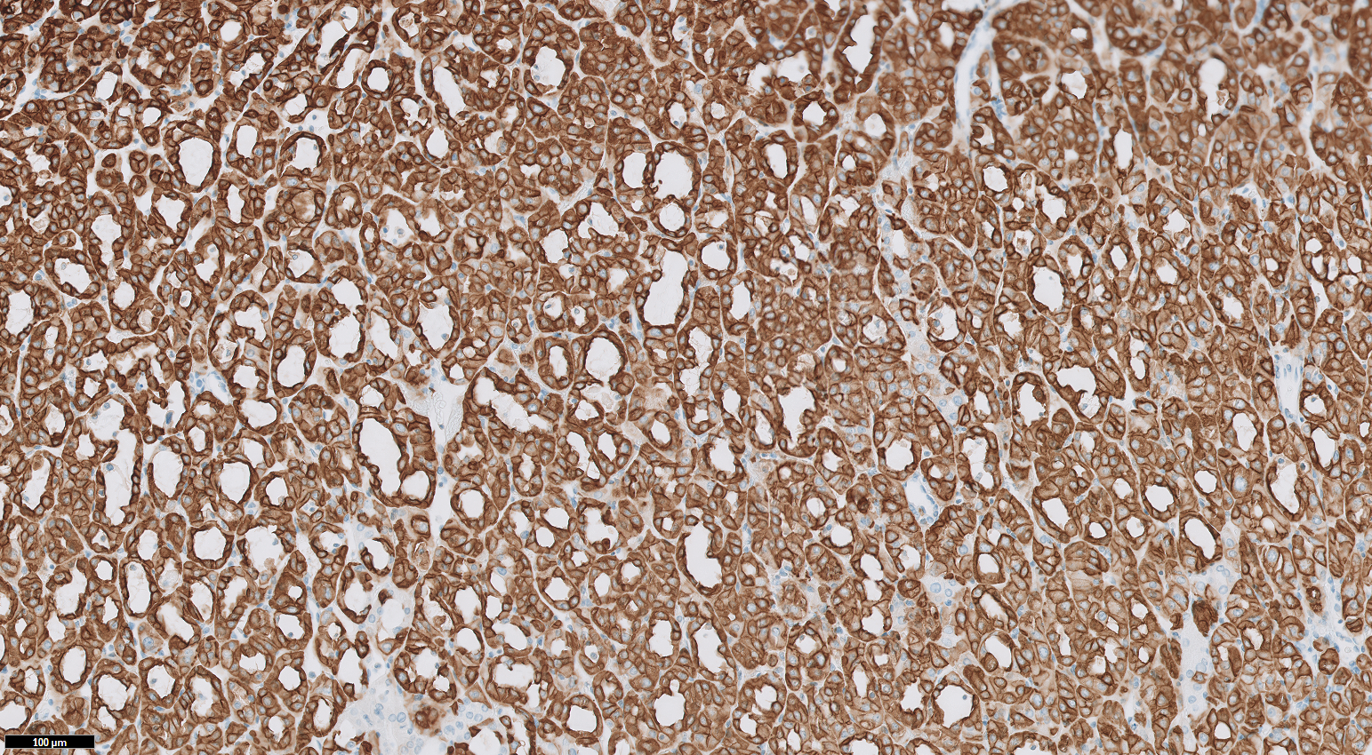
They present as relatively small, tan to brown tumours. LOTs are usually well circumbsribed unencapsulated lesions showing solid sheets and compact nests, with gradual transition to loosely connected strands with sharp delineation to oedematous stromal (aka ‘boats in a bay’ arrangement). The cells are eosinophilic, with bland round to ovoid (low-grade) nuclei lacking prominent nucleoli or nuclear irregularity. There often show perinuclear ‘halos’. They are characteristically diffusely positive for CK7 and negative for CD117 (1–4). The molecular data on these tumors are limited, with one study showing that LOTs are generally diploid tumours, sometimes with deletions of chromosomes 19p, 19q, and 1p(2) and another having identified TSC1/TSC2/mTOR mutations (5). To date, these neoplasms have not been found to have aggressive behaviour (hence the designation “tumour” is preferred to “carcinoma”) (1–5).
Key differential diagnosis
Oncocytoma: Different growth patterns including solid, nested, tubulocystic within abundant stroma (‘archipelaginous’ morphology). Small, central round to oval nuclei without perinuclear halos, prominent cell membranes, coagulative necrosis and papillary formations. CD117 (+) and Cathepsin K (-), CK7 (-) or only focally (+).
Oncocytic chromophobe RCC: More compact solid growth. The oncocytic cells have more pronounced cell membranes, raisinoid nuclei with irregular contours, binucleation, perinuclear halos. CD117 (+), MOC31 (+), diffuse CK7 (+), Cathepsin K (-).
EVT: Diffuse sheets, nests or tubulocystic growth pattern. The cells have abundant eosinophilic cytoplasm and marked intracytoplsmic vacuoles, “high-grade nuclei”, often with prominent nucleoli. Cathepsin K (+), CK7 (-), CD117 (+) , CD10 (+).
ESC RCC : Solid and cystic growth with characteristic cytoplasmic coarse or fine granular stippling (reminiscent of ‘leismaniasis’). These tumours usually show at least focally CK20 (+). CD117 (-), CK7 (-).
References
Summary of clinical history
62-year-old female with incidentally discovered left renal lesion on imaging underwent robotic partial nephrectomy.
Gross findings
Well defined nodule in the left kidney measuring 30 x 25 x 17 mm. Cream-tan cut surface.
Microscopic findings
CLICK IMAGE TO ENLARGE