Author: Theodorus H. Van der Kwast MD, PhD Department of Pathology, University Health Network, Toronto, ON
64 year-old man underwent radical prostatectomy for an ISUP grade 3 prostate cancer, incidental microscopic finding right mid anterior in anterior fibromuscular stroma
Gross findings
Well-circumscribed cellular nodule, approximately 7 mm in diameter
Microscopic findings
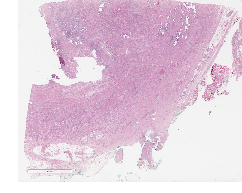
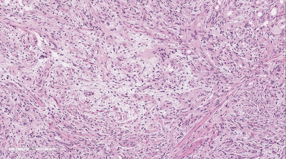
Well-circumscribed nodular area mid-anterior in the prostate (Figure 1)
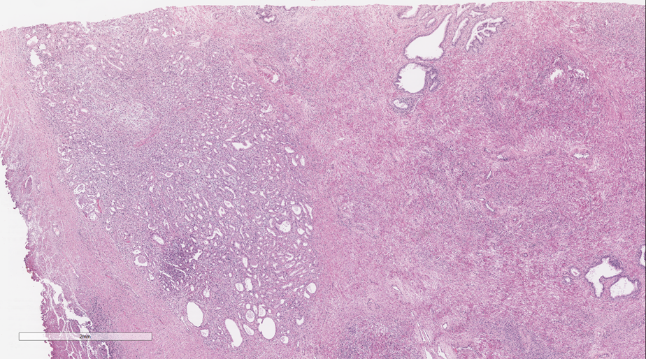
The nodule is composed of densely packed glands (Figure 2)
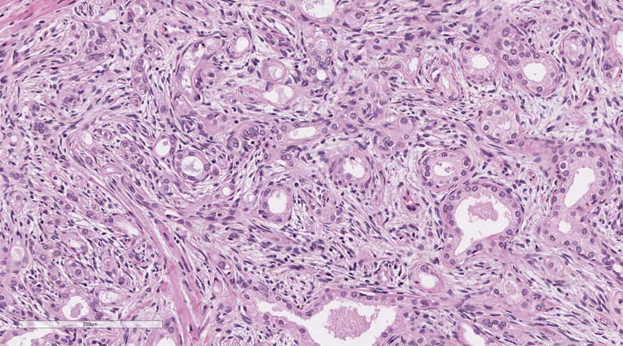
Cellular stroma and glands are closely admixed (Figure 3)
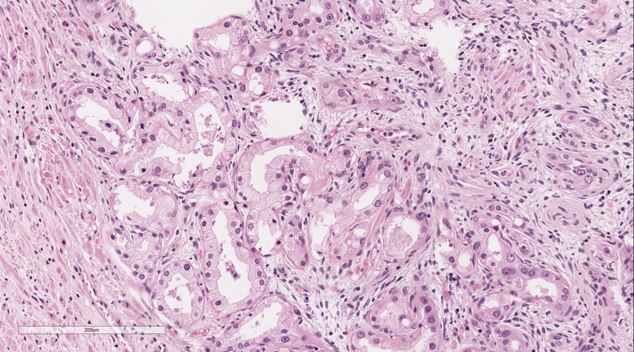
Glands are small-sized, have pale cytoplasm and somewhat enlarged nuclei with rather inconspicuous nucleoli (Figure 4)
Area with myxoid appearance is visible (Figure 5)
Immunohistochemical and Molecular findings
Positive:
PSA – glandular cells
HMWK
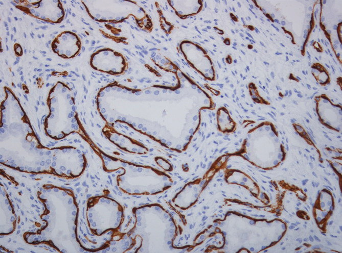
Smooth muscle actin (Figure 6)
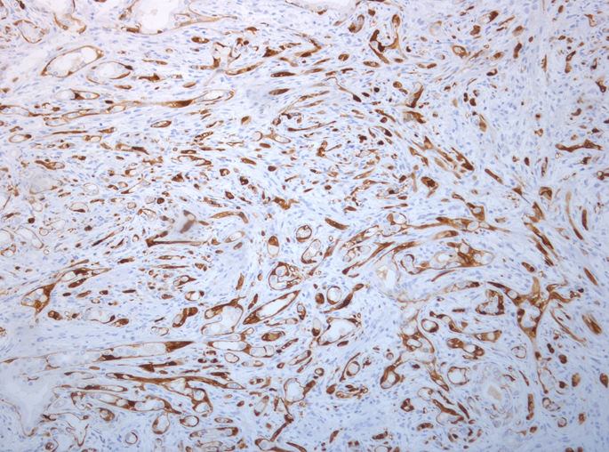
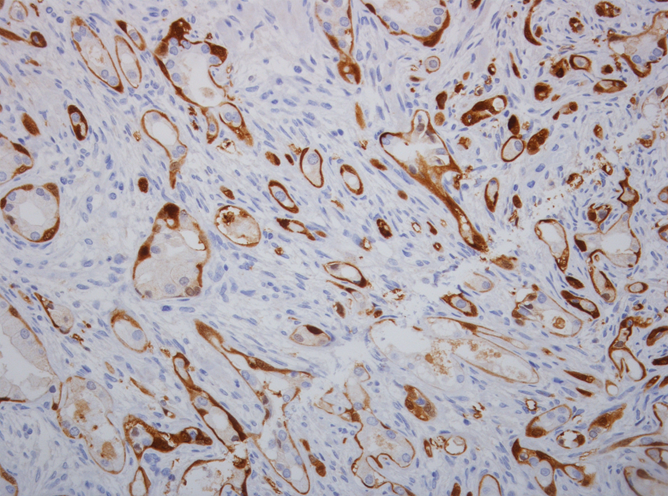
S100 (Figures 7 and 8)
Test yourself. What diagnosis would you give to this case?
Adenosis (atypical adenomatous hyperplasia)
Adenocarcinoma, Gleason grade 1
Nephrogenic remnants
Sclerosing adenosis
Blue nevus
Diagnosis by author:
Sclerosing adenosis
Discussion of case
Sclerosing adenosis is a benign lesion of the transition zone of the prostate, characterized by a cellular nodule of mostly small-sized glands and solid nests in a cellular spindle-cell stroma (1). Characteristically, the basal cell layer shows myoepithelial differentiation and is positive for S100, smooth muscle actin, and HMWK (2). Cytologic atypia is minimal, but rare cases may demonstrate nuclear atypia (3).
Major Differential Diagnosis
Adenosis (atypical adenomatous hyperplasia) Nodular growth, transition zone localization Sporadic basal cells lining small-sized (pale) glands Lacks intervening dense cellular stroma and myxoid areas
Adenocarcinoma No basal or myo-epithelial cells lining acini More conspicuous nucleoli
Nephrogenic remnants Lobule of small-sized uniform glands with characteristic eosinophilic secretion
Blue nevus Pigmented (S100 positive) stroma cells No glandular component
Key References
Young RH, Clement PB. Sclerosing adenosis of the prostate. Arch Pathol Lab Med. 1987; 111: 363-6.
Grignon DJ, Ro JY, Srigley JR, Troncoso P, Raymond AK, Ayala AG. Sclerosing adenosis of the prostate gland. A lesion showing myoepithelial differentiation. Am J Surg Pathol. 1992; 16: 383-91.
Cheng L, Bostwick DG. Atypical sclerosing adenosis of the prostate: a rare mimic of adenocarcinoma. Histopathology. 2010; 56: 627-31.